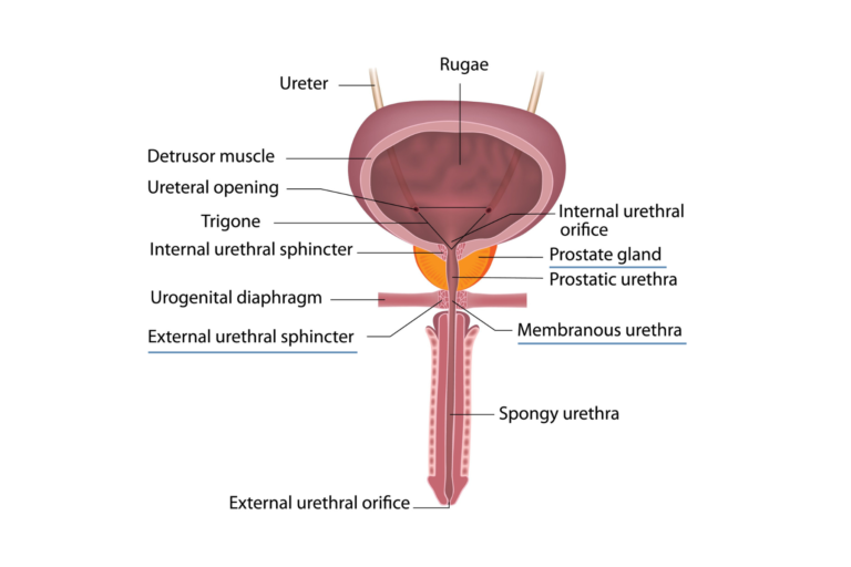
Male Pelvis Kayrote Biology Diagrams This MRI male pelvis axial cross sectional anatomy tool is absolutely free to use. Use the mouse scroll wheel to move the images up and down, or alternatively, use the tiny arrows (→) on both sides of the image to navigate through the images. For a more detailed view, double-click the image to view it in full screen, and use the menu in the PELVIC STRUCTURES. The relationships of male pelvic structures are illustrated in these complementary sagittal views—a paramedian and a median section. In the lower median view, the complete course of the urethra from the bladder to the meatus at the end of the penis, its passage through the prostate gland and the urogenital diaphragm, is shown.
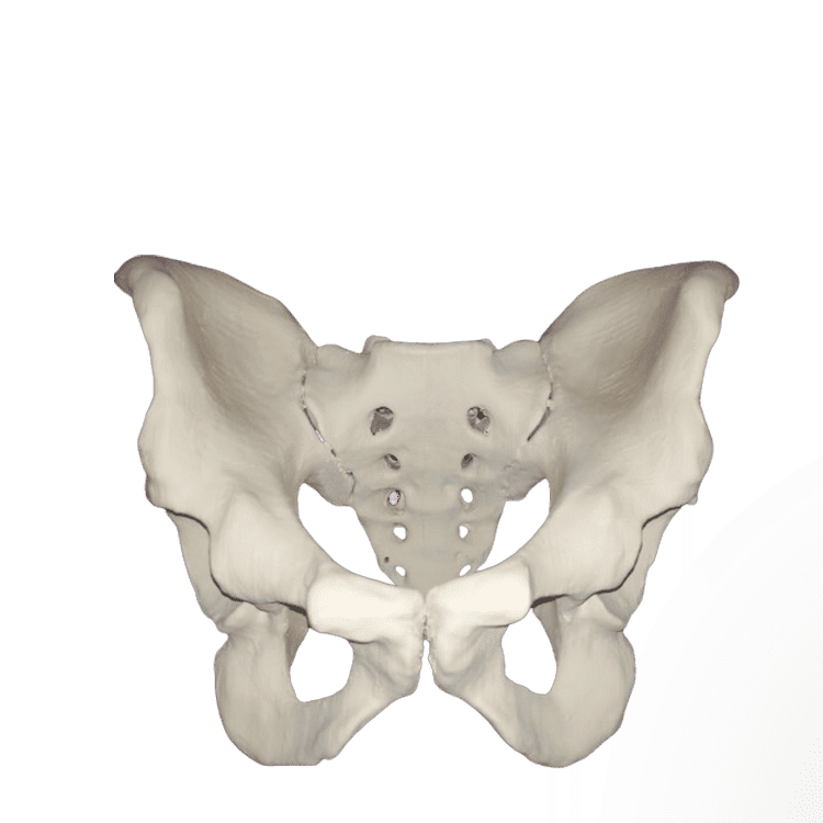
Learn about the bones, joints and shapes of your pelvis, the bony structure inside your hips, buttocks and pubic region. Find out how your pelvis differs by sex and what conditions can affect it.
Atlas of anatomy of the male pelvis and reproductive system Biology Diagrams
22.6: MODELS- Male Hemi-Pelvis and Torso This page provides a detailed overview of the male reproductive system's anatomy and histology, covering structures like the scrotum, testes, ejaculatory duct, urethra, and penis, along with components such as the dartos muscle and seminal vesicles.
Learn about the anatomy of the male pelvis, including prostate, bladder, genital organs, rectum and more. See MRI images and labels of the pelvic structures and their zones, regions and fascia.

Free Male Pelvis Axial Anatomy - mrimaster Biology Diagrams
This e-Anatomy module contains seventy-nine illustrations dedicated to the anatomy of the male pelvis and genital system. These fully annotated anatomical illustrations are presented as a comprehensive atlas of the male reproductive system, specifically designed for medical students, residents and healthcare professionals. Material and methods